Radiographic Examination of the Ankle Bones
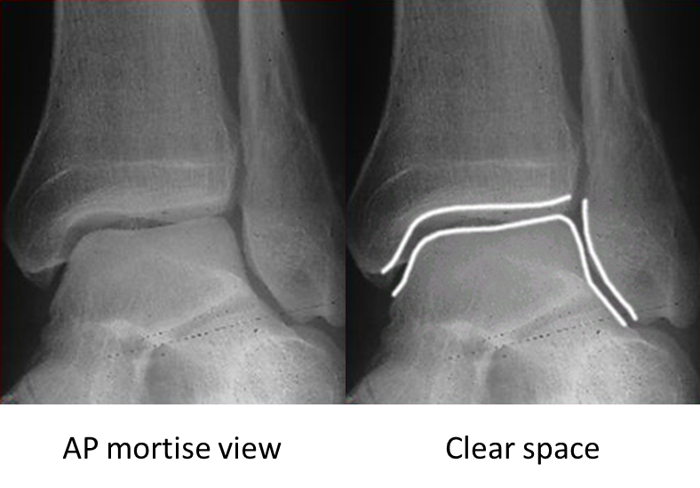
AP mortise view
The AP mortise view is done with the leg internally rotated 15-20o so that the x-ray beam is perpendicular to the inter-malleolar line. This view permits examination of the articular space (clear space). The width of the clear space between the talus and the articular surfaces of the medial malleolus, the tibial plafond and the lateral malleolus should be equal.