Carpal Bones
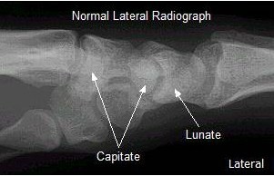
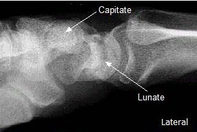
Step 6: The lateral arcs
On the lateral view, the opposing joint margins of the capitate, lunate, and radius should form four parallel arcs. In addition to the line through the radius discussed in step 5, you should also look for disruptions of these arcs. Disruption of these arcs is another marker for dislocation of one of the carpal bones. Note: on the lateral radiograph furthest to the right, the joint margin of the capitate is not aligned with the joint margin of the lunate. This is called a perilunate dislocation of the capitate.
 |
.JPG) |
 |