Digital Rectal Exam & Anoscop
Anoscopy
- Lubricate the anoscope well with the obturator in place
- Spread the buttocks and gently insert the anoscope (with obturator) into the anal canal
- Asking the patient to take a few deep, gentle breaths and to bear down slightly may make the insertion easier
- Gently advance the instrument towards the umbilicus until the full length is inserted
- If the patient complains of pain during insertion, note the location and quality and correlate the pain with clinical symptoms
- Remove the obturator and visualize the anal mucosa
- Any fecal matter can be removed with a large swab
- Note the gross appearance of mucous membranes and vasculature as well as the presence of pus, mucous, blood, ulceration, and hemorrhoidal tissue
- Slowly rotate the anoscope (with the obturator still removed) as it is withdrawn, inspect the anal canal as the device is extracted, looking for mass lesions, hemorrhoids or fissures. A clear plastic anoscope allows the examiner to visualize the mucosa both through the walls of the anoscope as well as at the opening of the device
- Masses or polyps visible through the anoscope should not be sampled as the instrument is too short to get a good appreciation of the extent of the mass lesion noted. A sigmoidoscope, either rigid or flexible is best used here.
- Once the device is fully withdrawn from the patient's anal canal, wipe away any excess lubricant with tissue
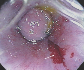
Anal fissure (arrow) seen through the wall of a clear plastic anoscope