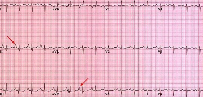
Right atrial enlargement
Patients presenting with RAE demonstrate an ECG pattern in which the P wave duration is unaffected, but its shape is peaked and its amplitude is increased to greater than 2.5 mm in leads II, III, aVF (arrows below) and sometimes V1. With extreme enlargement of the right atrium, the P wave may demonstrate terminal negativity in lead V1, resembling LAE. Right atrial enlargement is commonly associated with congenital heart disease, tricuspid valve disease, pulmonary hypertension and diffuse lung disease. Furthermore, patients presenting with RAE often demonstrate ECG changes associated with right ventricular hypertrophy as well.